Guide
The science of melanin: how skin makes pigment
A plain-language look at how pigment-making cells build melanin with tyrosinase and pass it to the skin cells around them.
On this page
Where skin pigment is made
The color of skin, hair, and eyes comes from a pigment called melanin, and nearly all of it is produced by a specialized cell called the melanocyte. Melanocytes originate early in development from the neural crest and then migrate outward, settling in the deepest layer of the epidermis (the basal layer), in hair follicles, and in tissues such as the eye and the inner ear. In the skin, they sit at the boundary between the epidermis and the layers beneath, positioned to distribute pigment upward toward the surface.
Inside each melanocyte, melanin is not made loose in the cell. It is built and stored inside dedicated compartments called melanosomes, which are membrane-bound organelles related to lysosomes. Researchers describe melanosomes as maturing through recognizable stages: they begin as structural scaffolds with little pigment and gradually fill with melanin until they are densely packed, pigment-laden granules. This staging keeps the chemistry of pigment production contained, because several of the intermediates involved are reactive.
Melanocytes are far outnumbered by the surrounding keratinocytes, the main structural cells of the epidermis. A single melanocyte extends slender, branch-like projections that reach many neighboring keratinocytes at once. This arrangement matters, because a melanocyte's job is understood to be less about coloring itself and more about manufacturing pigment and distributing it to the cells around it. This article is educational and describes how the process is currently understood; it is not medical advice.
Tyrosinase, the enzyme at the center of the process
The chemistry of melanin production is driven by a copper-containing enzyme called tyrosinase, which is widely described as the rate-limiting step of pigment synthesis — the point that sets the overall pace. Tyrosinase acts on the amino acid L-tyrosine, and this is where the pathway that gives the pigment its name begins. Because tyrosinase controls the tempo, the amount of active enzyme a melanocyte carries is one of the central factors in how much pigment it can make.
Tyrosinase is understood to catalyze the first two steps in a linked sequence. It first helps convert L-tyrosine into a compound called L-DOPA (levodopa, or 3,4-dihydroxyphenylalanine), and then oxidizes L-DOPA into dopaquinone. Dopaquinone is the pivotal molecule of the whole pathway: it is highly reactive, and the direction it takes from here determines which kind of melanin the cell ultimately produces. Everything upstream funnels into this single, branching intermediate.
Tyrosinase does not work alone. Melanocytes also carry related proteins, including tyrosinase-related protein 1 (TYRP1) and dopachrome tautomerase (DCT, also called TYRP2), which are understood to shape later steps and influence the final pigment. The production of tyrosinase and its partners is governed largely by a master control protein called MITF (microphthalmia-associated transcription factor), which switches on the genes for the pigment-making machinery when the cell receives the right signals.
Eumelanin and pheomelanin: two pigments, one fork in the road
There is not one melanin but two broad families, and the difference between them is set at the dopaquinone branch point. When dopaquinone continues down a pathway that runs through indole-type intermediates (such as DHI and DHICA), it forms eumelanin — the brown-to-black pigment. When dopaquinone instead encounters sulfur-containing molecules, chiefly the amino acid cysteine (supplied in part through glutathione metabolism), it is diverted to form pheomelanin — the reddish-to-yellow pigment. The same starting enzyme and the same key intermediate can therefore lead to strikingly different colors.
In practice, human skin and hair contain a blend of both pigments rather than one alone, and it is the ratio between them, along with the total quantity, that research associates with the range of natural coloring people have. A shift toward eumelanin is associated with darker, browner tones; a shift toward pheomelanin is associated with lighter, redder tones, including the coloring often seen with red hair and fair skin. The mix is not fixed at a single value but reflects the balance of conditions inside the melanosome.
The two pigments are also understood to differ in how they behave under ultraviolet (UV) light. Eumelanin is generally described in the research literature as the more effective absorber of UV radiation and the more photoprotective of the two, while pheomelanin is studied differently in this respect. These are descriptions of the pigments' physical properties as understood by researchers, not statements about any product, treatment, or health outcome.
Handing pigment to neighboring skin cells
Making melanin is only half of the story; the pigment then has to be delivered to the cells that carry it toward the surface. This is the purpose of the melanocyte's branch-like dendrites. As melanosomes mature and fill with pigment, they are transported outward along these projections toward their tips, positioning the finished granules where they can be passed to surrounding keratinocytes. The functional partnership of one melanocyte supplying pigment to its cluster of neighboring keratinocytes is often called the epidermal melanin unit.
Exactly how a melanosome crosses from one cell into another is an area of active study, and several mechanisms have been proposed and described. These include:
- The keratinocyte engulfing the pigment-tipped end of a dendrite
- The release of pigment packets that the neighboring cell then takes up
- Direct membrane-to-membrane transfer
Researchers do not treat these as mutually exclusive; the current picture is that pigment hand-off may occur through more than one route.
Once melanosomes are inside a keratinocyte, they do not scatter randomly. They are frequently observed to gather on the sun-facing side of the cell's nucleus, forming a cap-like arrangement over it. This positioning is understood to place pigment between incoming UV light and the cell's DNA, which is why melanin is so often described in terms of a light-absorbing, shielding role. As keratinocytes mature and rise toward the skin surface, they carry this pigment with them, which is what makes the color visible.
What tells a melanocyte to make more
Pigment production is responsive, not static, and one of the best-studied signals is ultraviolet light. When skin is exposed to UV, keratinocytes are understood to respond in part through the protein p53 and to release signaling molecules — including alpha-melanocyte-stimulating hormone (α-MSH), derived from a precursor called POMC. These messengers travel to nearby melanocytes and bind a receptor on their surface called the melanocortin 1 receptor (MC1R). This receptor-and-messenger relay is the mechanism most commonly used to explain the delayed tanning that follows sun exposure.
When α-MSH activates MC1R, it raises the level of an internal messenger called cyclic AMP (cAMP) inside the melanocyte, which in turn activates MITF — the same master regulator described earlier. MITF then increases production of tyrosinase and its partner proteins, and the cell's overall pigment output rises. Naturally occurring variants in the MC1R gene are understood to change how strongly this receptor responds, and such variants are associated with a shift toward pheomelanin and with red-hair, fair-skin coloring.
An educational note on melanin science
This article describes how melanin production is currently understood at the level of cells, enzymes, and signaling pathways. It is educational content, not medical advice, and it does not diagnose any condition, recommend any treatment, or tell you what is appropriate for your skin. Pigmentation is influenced by genetics, sun exposure, hormones, and many other factors, and questions about your individual skin belong with a licensed provider who can consider your full situation.
Some prescription ingredients are studied in the context of skin pigmentation and cell turnover, and researchers describe various points in the pathways above — the activity of tyrosinase, the maturation of melanosomes, and the transfer of pigment to keratinocytes — as targets of scientific interest. Describing a mechanism is not a claim that any product produces a particular result. Where compounded medications are involved, it is important to know that compounded preparations are not FDA-approved products, and statements about them have not been evaluated by the FDA.
On a telehealth service, no medication is prescribed on the basis of an article. An independent, licensed provider reviews a person's medical intake and makes every clinical decision. The purpose of a guide like this one is only to make the underlying biology clearer, so that any later conversation with a qualified clinician starts from a shared, accurate understanding of how the skin builds and shares its pigment.
Common questions
- What is melanin, and what is it made from?
- Melanin is the pigment that gives skin, hair, and eyes their color. It is built inside pigment-making cells called melanocytes, starting from the amino acid L-tyrosine. The enzyme tyrosinase converts tyrosine into a series of reactive intermediates that are assembled into melanin and stored in compartments called melanosomes.
- What does tyrosinase do in the pigment pathway?
- Tyrosinase is a copper-containing enzyme widely described as the rate-limiting, pace-setting step of melanin production. It helps convert L-tyrosine into L-DOPA and then into dopaquinone, the pivotal intermediate whose fate determines which type of melanin the cell makes. How much active tyrosinase a melanocyte carries strongly influences how much pigment it can produce.
- What is the difference between eumelanin and pheomelanin?
- They are the two broad families of melanin, and the split happens at the dopaquinone step. Eumelanin is the brown-to-black pigment formed when the pathway continues through indole-type intermediates; pheomelanin is the reddish-to-yellow pigment formed when sulfur-containing cysteine diverts the pathway. Human coloring reflects a blend of both, and the ratio between them is associated with the range of natural tones, including red-hair and fair-skin coloring.
- Do people with darker skin have more melanocytes?
- Generally no. Research indicates that the number of melanocytes is broadly similar across populations. The differences in skin color are attributed mainly to how melanosomes are made and handled — their size and number, how much and which type of melanin they contain, and how they are distributed and broken down within the surrounding keratinocytes.
Ready when you are
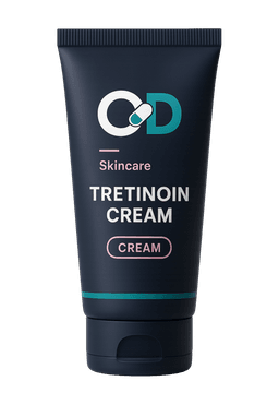
Tretinoin Creamfrom $79.00
- Your exact strength
- Licensed provider review
- Full refund if declined
Browse skincare & dermatology
Exact strengths and prices up front — reviewed by a licensed U.S. provider.
This guide is for general education and is not medical advice. Compounded medications are not FDA-approved drugs, and statements on this site have not been evaluated by the FDA. A licensed provider reviews every prescription request.